Synthetic Generation Pipeline
To photorealistic microscopy images in 5 steps.
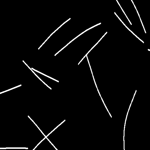
1
Generating Geometry
Generate MT masks with realistic geometry.
→
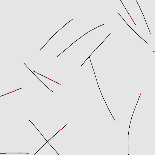
2.1
Physical Rendering
Apply optical system simulation.
→
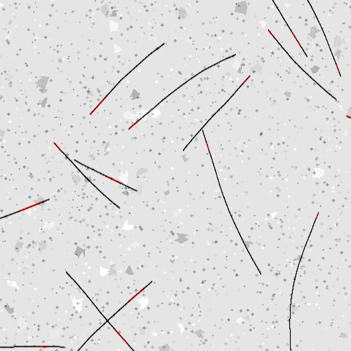
2.2
Artifact Simulation
Add realistic imaging artifacts.
→

2.3
Noise Addition
Inject sensor and shot noise.
→
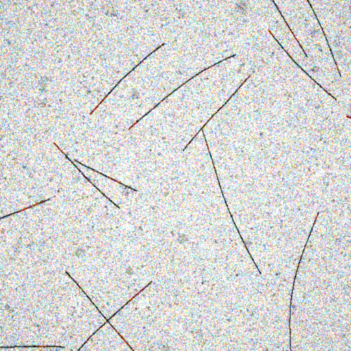
2.4
Global Distortions
Apply intensity and contrast variations.
Optimizing θ aligns synthetic image distributions with real microscopy data without the need for annotations.
